
Study: Clinical Benefit of AI-Detected Breast Cancer Remains Uncertain
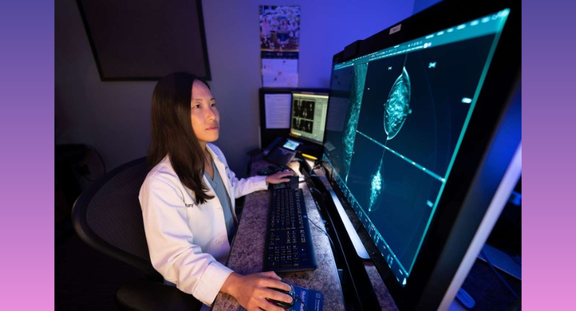
Artificial intelligence (AI) may help radiologists find small signs of breast cancer that are missed during regular mammograms. It may also find patterns linked to a higher risk of cancer before tumors can be seen. However, important limits remain, according to a review led by researchers at the UCLA Health Jonsson Comprehensive Cancer Center.
Published in the Journal of Breast Imaging, the review looks at AI tools currently sold for helping doctors read screening mammograms. It also examines research into whether these tools can find or predict interval breast cancers.
Interval cancers are breast cancers found after a woman has a normal screening mammogram but before her next planned screening. They are an important way to measure how well a breast cancer screening program works. These cancers are often more aggressive than cancers found during regular screening.
“The main goal of screening mammograms is to prevent these interval cancers and find as many cancers as possible earlier, during screening,” said Tiffany Yu, M.D., an assistant professor of radiology at the David Geffen School of Medicine at UCLA and the study's senior author. “As AI tools become more widely available, we wanted to give readers a timely overview and a basic understanding of interval cancers and how AI might help find them. Making sure these tools are safe and work well for patients is very important.”
Some interval cancers grow quickly and cannot be seen on the previous mammogram. Others show small signs on the earlier mammogram that were not noticed during screening. AI may help radiologists find cancers earlier in this second group.
The researchers reviewed studies of two possible uses for AI. First, they looked at whether AI could find interval cancers that could be seen, in hindsight, on an earlier mammogram. Second, they looked at whether AI could find small patterns on mammograms that looked normal but were linked to a higher chance of developing breast cancer later.
In studies that looked back at earlier mammograms, AI found a large share of interval cancers that had small visible signs. The estimates varied greatly, from about 5% to 78%. The results depended on the study, the AI system and the methods used.
However, the researchers stress that finding possible signs of cancer in past images does not prove that AI lowers the number of interval cancers. Most studies looked back at old records. Researchers examined mammograms taken before women were diagnosed with breast cancer to see whether AI could have spotted signs of the disease.
“Finding a cancer by looking back at old images is very different from showing that using AI during regular screening would have led to an earlier diagnosis,” said Hannah Milch, M.D., an associate professor of radiology at the David Geffen School of Medicine at UCLA and a co-author of the study. “We need studies that follow patients over time and show that AI actually improves their health outcomes.”
Some studies also suggest that AI may identify women with a higher risk of breast cancer even when their mammograms look normal. In one study, AI gave its highest risk scores to 23.1% of women who later developed interval cancer, three screening rounds before their diagnosis. That share rose to 39.4% on the mammogram taken just before diagnosis.
These findings suggest that AI might warn doctors about cancer risk before a cancer can be seen. But doctors do not yet know what they should do when AI gives a woman a high risk score and her mammogram shows no signs of cancer. More imaging might find cancer earlier, but it could also lead to false alarms, unnecessary medical procedures and overdiagnosis.
The review also discusses a large randomized controlled trial from 2026. The study included more than 105,000 women and compared screening supported by AI with standard double reading. Double reading is a common practice in Europe in which two radiologists examine the same mammogram. The rate of interval cancers was 1.55 per 1,000 women in the AI-supported group, compared with 1.76 per 1,000 in the standard screening group. AI-supported screening also had higher sensitivity, at 80.5% compared with 73.8%. Both groups had the same specificity of 98.5%.
The AI-supported method reduced the screening workload by 44.3% without lowering the overall rate of cancer detection.
The authors say this trial provides some of the strongest evidence so far from a study that followed patients forward in time. However, they warn that the trial was designed to show that AI-supported screening was not worse than standard screening, not to prove that it lowers interval cancer rates. The screening approach also differed from many U.S. programs. It used 2D mammograms, longer gaps between screenings and two radiologists reading each mammogram.
More generally, the researchers found major differences between studies of AI and interval cancers. Studies use different definitions of interval cancer and different screening technologies, time gaps between screenings and ways of assigning work to radiologists. Most studies also test only a small number of AI systems. This makes it hard to know whether results from one system also apply to others.
“These differences make it hard to compare results directly because screening programs vary between countries, and there are few studies that apply to U.S. programs,” Yu said. “This also shows where more research is needed before these tools can become a regular part of patient care.”
The researchers say future studies should follow patients over time and test whether AI-supported screening lowers interval cancer rates. They should also examine how it affects the number of women called back for more tests, false alarms, radiologists' workloads and follow-up testing. Long-term patient health and ongoing checks of AI systems after they become available for sale will also be important for understanding how safe and effective they are.
The study's first author is Javier Galvan, M.D., from the Department of Radiological Sciences at UCLA. The other author is Niki Nourmohammadi, D.O., a radiology resident at Larkin Health System.